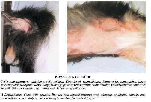
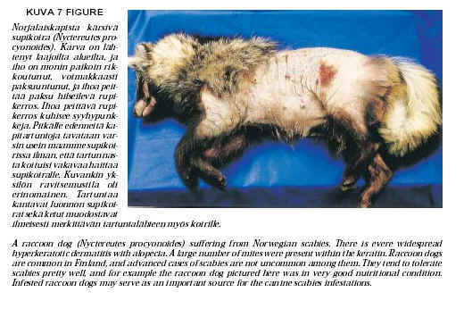
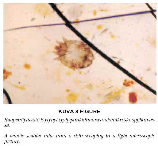
Canine scabies, sarcoptic mange is caused by an epidermal dwelling mite, Sarcoptes scabiei. The dogs are usually infested by the canine variant of the mite, S. scabiei var. canis. Wild raccoon dogs (Nyctereutes procyonoides) and foxes (Vulpes vulpes) are commonly infested by S. scabiei var. vulpes in Finland, and these species serve as a source of scabies for dogs as well. Canine scabies is characterized by intense pruritus and the skin lesions, which are usually present on the ear margins and on the ventral trunk. The disease is difficult to diagnose, as it is not easy to find mites, and as the disease resembles other pruritic dermatoses. Nowadays, the diagnosis can be based on the demonstration of specific serum antibodies to S. scabiei. The use of avermectins and milbemycins has more and more substituted for the arduous treatments with dips with hair clippings. Canine scabies is a zoonosis. This article reviews the current information about the morphology (presented with scanning electron micrographs) and the biology of the scabies mite, as well as the epidemiology, clinical features, diagnosis and treatment of canine scabies.
Rupture of the common digital extensor tendon in neonatal foals is characterized by fluctuating swelling over the dorsolateral aspect of the carpus. When moving, affected foals knuckle over at the fetlock. A distinct syndrome of congenital hypothyroidism and dysmaturity recognized in North America, manifests as rupture of the common digital extensor tendon(s), flexural deformities, mandibular prognathia and incomplete skeletal ossification. This retrospective study was carried out by investigating the 1986–1999 records of the Large Animal Clinic, University of Helsinki. The aim of the study was to examine the incidence, treatment options and prognosis of ruptured extensor tendons in foals, and to establish whether this congenital hypothyroidism and dysmaturity syndrome also occurs in Finland.
Fourteen foals presenting with clinical signs consistent with rupture of the common digital extensor tendon were included. All foals were younger than two weeks when the rupture was diagnosed. The rupture was bilateral in eight and unilateral in six foals. Seven foals had flexural deformities, most commonly in carpus. Incomplete skeletal ossification was diagnosed in seven foals. One foal exhibited a combination of four lesions consistent with the congenital hypothyroidism and dysmaturity syndrome; five foals had three lesions, respectively.
All foals had stall rest for 1–4 weeks combined with supportive bandaging (except one foal), and in some foals bandages with splints were used. Restricted exercise was allowed after clinical signs subsided. Foals with a ruptured digital extensor tendon as the only lesion recovered well. In foals with other musculoskeletal lesions, the prognosis was guarded and was dependent on the severity of the other lesions. Four of the foals have been euthanized for reasons unrelated to the tendon rupture. A thorough physical inspection is warranted in foals with ruptured extensor tendons to rule out a variety of other related, more severe musculoskeletal lesions.