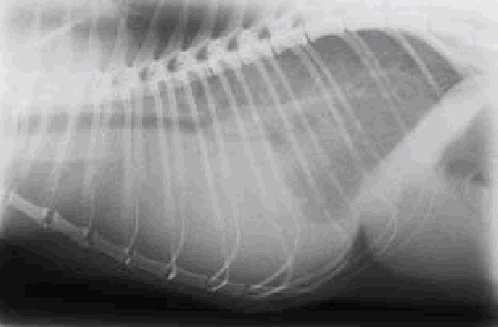
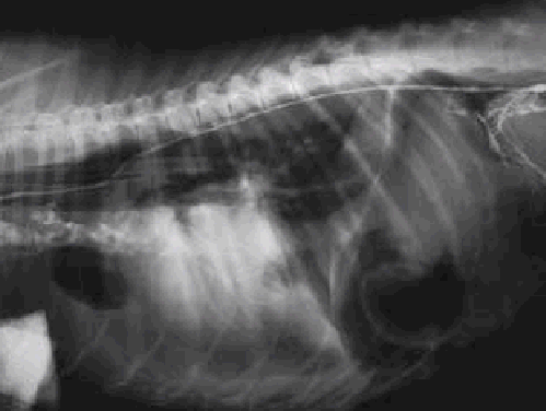
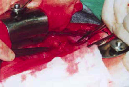
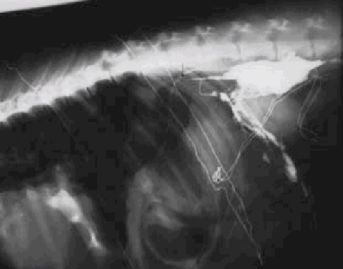
Idiopathic chylothorax is the accumulation of chyle in the pleural cavity causing anorexia, coughing, respiratory distress and exercise intolerance. Diagnosis is made by taking radiographs, blood samples and an effusion sample. An effective medication has not been found. The goal of the treatment is to decrease the accumulation of chyle and improve the resorption to the systemic circulation. It is possible to obtain a fairly good result with a diet, rutin medication and surgery. Prognosis is still guarded or poor.
This report describes the diagnostics and treatment of chylothorax in the dog and the cat along with a typical canine patient developing chylothorax despite of surgery. No explanation to the symptoms was found by postmortem examination.
A health management project in dairy cattle herds was carried out in 1998 and 1999 in Southern Ostrobotnia, Finland, by Cooperative Maitojaloste. This article covers information about 95 dairy farms, which participated in the project. The farms were visited twice at a yearly interval.
The purpose of this study was to specify the health state of the farms in the first year and the benefit from the health management work after one year. Farm data was collected from the latest herd production and health reports, by interviewing the producers and from observations made by the veterinarians and dairy�s production advisers.
The herd health was charted and health management plans were made during the farm visits. The targets for the different sectors of health management were specified on the grounds of these visits. The sectors were udder health, foot health, parasite problems, calf health, reproduction, nutrition, infectious diseases, animal drug residues and housing conditions.
In the year 1998 fourteen farms, and in the follow-up year 42 farms, reached the health goals of all sectors. The number of health problems in different sectors decreased on 56 farms, remained the same on 20 farms and increased on 10 farms. The data collected at the farms was estimated statistically. The increase in the average milk production, the decrease in the udder inflammation percentage, and the decrease in the use of antibiotic treatment for mastitis were statistically significant.
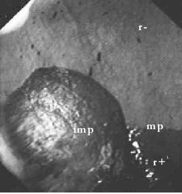
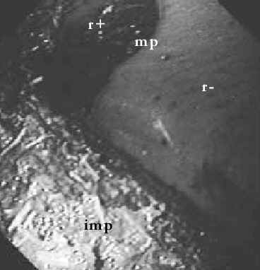
In gastric impaction of horse, dry, solid food material accumulates in the stomach. The most reliable method for diagnosing gastric impaction is gastroscopy, although a 3-metre-long endoscope is needed for adult horses.
Presence of solid material in the stomach after routine fasting prior to endoscopy (24 hours) can be considered abnormal. Various extrinsic and intrinsic aetiological factors have been described, but often the underlying cause remains unknown. Gastric ulceration may also be present in horses with gastric impaction. Gastric impaction may cause varying symptoms, such as intermittent low-grade colic or inappetence, or the horse may be relatively asymptomatic. Horses with gastric impaction generally have normal findings at routine physical examination, even though slight dehydration may be present. Ancillary laboratory work is usually of no benefit.
Conservative treatment consists of withholding food, hydrating the mass via nasogastric tube, and prokinetic drugs to stimulate gastric motility. Most cases respond to conservative treatment within a couple of days. In unresponsive and chronic cases surgical treatment may be considered. At surgery, saline is injected through the stomach wall into the impaction mass, and additional softening can be obtained with massage of the mass. If treatment is unsuccessful, gastric impaction may result in damage, including rupture, of the gastric wall. The prognosis of chronic impaction is guarded.